FIGURE 2:
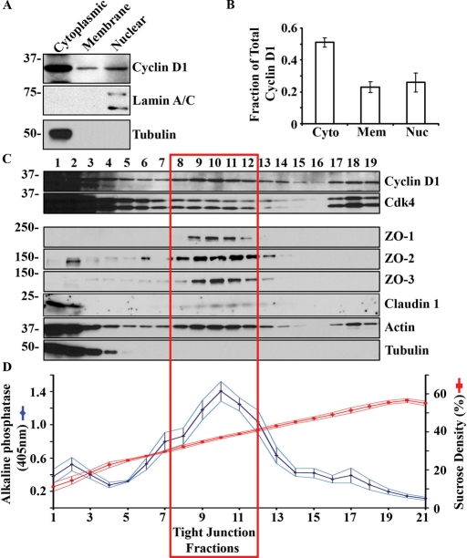
Cyclin D1 cosediments with TJs in polarized epithelial cells. (A) Representative Western blot of cyclin D1 in isolated SKCO-15 cytosolic, cell membrane, and nuclear fractions. Tubulin is shown as a marker of cytosolic protein and lamin A/C as a nuclear marker. (B) Densitometry analysis of Western blot; n = 3. (C) Sucrose density gradient fractionation of SKCO-15 cell lysate showing cosedimentation of cyclin D1 with tight junction proteins. (D) Sucrose density of indicated fractions (15–60%; red) and ALP activity in the corresponding fractions (blue; n = 8).

