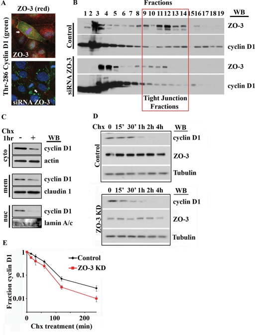
FIGURE 6:
ZO-3 is required for cyclin D1 TJ localization and cyclin D1 protein stability. (A) SKCO-15 cells treated with siRNA directed against ZO-2 (top panel) or ZO-3 (bottom panel) and immunostained for Thr-286 cyclin D1 (green) and ZO proteins (red). Arrows highlight mitotic cells. (B) Sucrose density gradient of colonic epithelial cells after ZO-3 depletion. Cyclin D1–ZO-3 cosedimentation in TJ fractions is diminished after ZO-3 depletion. (C) Subcellular fractionation reveals differential cyclin D1 protein stability after Chx treatment (50 μM, 1 h). (D) Cyclin D1 shows lower protein stability in ZO-3 siRNA-treated cultures. SKCO-15 cell lysates were harvested at the indicated times up to 4 h posttreatment with Chx (50 μM). (B) Densitometric analysis of Western blot data shows cyclin D1 protein levels in SKCO-15 cells after Chx treatment (n = 3).

