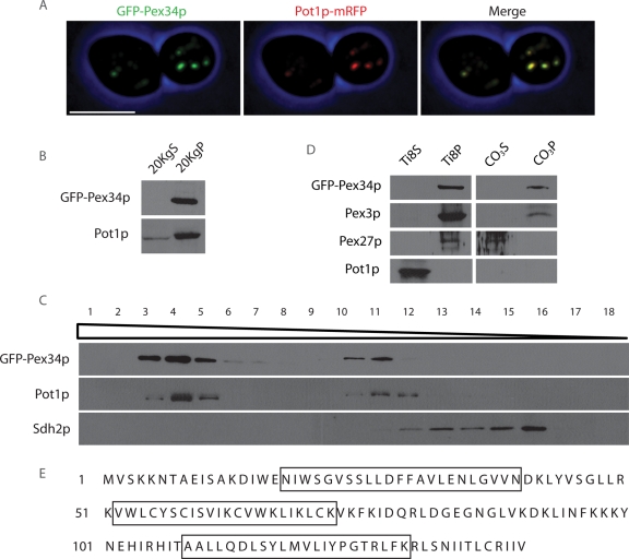
FIGURE 1:
Pex34p is a peroxisomal integral membrane protein. (A) GFP-Pex34p colocalizes with the chimeric peroxisomal marker protein Pot1p-mRFP to punctate structures characteristic of peroxisomes by confocal fluorescence microscopy. Bar, 5 μm. (B) GFP-Pex34p localizes to the 20KgP subcellular fraction enriched for peroxisomes. Immunoblot analysis of equivalent portions of the 20KgS and 20KgP fractions from cells expressing GFP-Pex34p was performed with antibodies to GFP and to the peroxisomal matrix protein, Pot1p. (C) GFP-Pex34p cofractionates with peroxisomes. Organelles in the 20KgP fraction were separated by isopycnic centrifugation on a discontinuous Nycodenz gradient. Fractions were collected from the bottom of the gradient, and equal portions of each fraction were analyzed by immunoblotting. Fractions enriched for peroxisomes and mitochondria were identified by immunodetection of Pot1p and Sdh2p, respectively. (D) The 20KgP fraction from cells expressing GFP-Pex34p was treated with 10 mM Tris-HCl, pH 8.0, to lyse peroxisomes and was then subjected to ultracentrifugation to yield a supernatant (Ti8S) fraction enriched for matrix proteins and a pellet (Ti8P) fraction enriched for membrane proteins. The Ti8P fraction was treated further with 0.1 M Na2CO3, pH 11.3, and separated by ultracentrifugation into a supernatant (CO3S) fraction enriched for peripheral membrane proteins and a pellet (CO3P) fraction enriched for integral membrane proteins. Equal portions of each fraction were analyzed by immunoblotting with antibodies to GFP, the matrix protein Pot1p, the peroxisomal integral membrane protein Pex3p, and the peroxisomal peripheral membrane protein Pex27p. (E) Amino acid sequence of Pex34p. Boxed sequences designate three membrane-spanning regions predicted by SOSUI.

