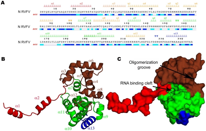
Figure 3. Structure of the N monomer.
(A) Sequence of the RVFV N protein with secondary structure elements indicated above. The colors correspond to those used to color the different sub-domains in the crystal structure of N shown in panels B and C. (B) Ribbon representation of the crystal structure of the RVFV N protein showing that the N terminus forms an arm (red) that extends from the globular core domain (brown and green). The C terminus, which is not involved in RNA binding, is shown in blue. The α-helices are labeled. (C) View of the RVFV N protein in surface representation. The orientation and color code are the same as in panel B.

