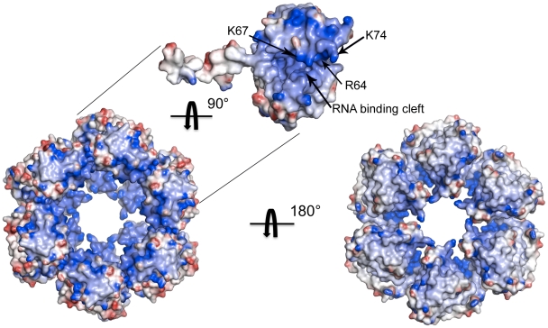
Figure 5. Electrostatic surface potential of the N hexamer.
Mapping of the electrostatic surface potential, from −10 kT in red to+10 kT in blue, onto the surface of hexamer I formed by native N protein reveals a patch of positive charges in the inner part of the ring, which likely accommodates the vRNA. Key residues in the RNA binding site are labeled on the electrostatic surface of a single monomer.

