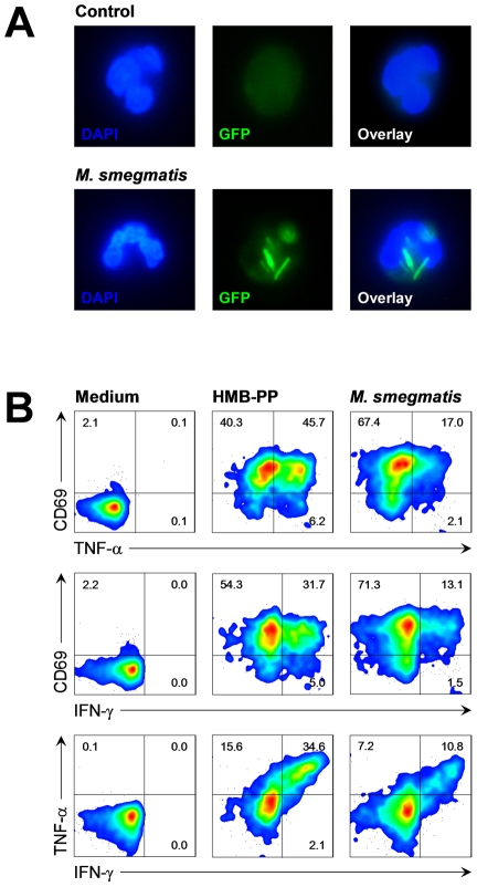
Figure 4. Vγ9/Vδ2 T cells respond to bacteria upon phagocytosis by neutrophils.
(A) Resting neutrophils or neutrophils after phagocytosis of M. smegmatis-gfp + stably expressing GFP, at a multiplicity of infection (MOI) of 10. Cells were counter-stained with DAPI and imaged by fluorescence microscopy. Data shown are representative from independent experiments using two different donors. (B) Activation of Vγ9/Vδ2 T cells co-cultured for 20 hours with neutrophils in the absence (medium) or in the presence of 10 nM HMB-PP, or co-cultured with neutrophils harboring M. smegmatis-gcpE + overexpressing HMB-PP synthase. Data shown are representative from independent experiments using three different donors.

