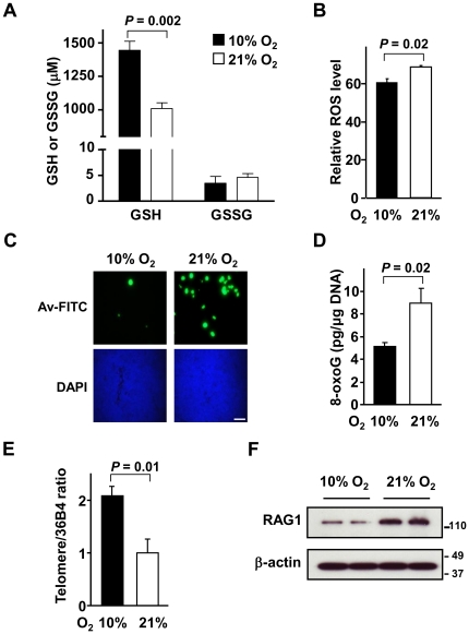
Figure 2. Lowering oxygen exposure reduces oxidative stress, DNA damage and genomic instability in thymus.
A. Changes in reduced (GSH) and oxidized (GSSG) glutathione levels show increased antioxidant capacity in blood of mice after chronic adaptation to the 10% oxygen condition. Data shown as mean ± SEM, with n = 4. B. Decreased ROS levels measured by DCF FACS in cells isolated from the thymus of mice in 10% oxygen. Data shown as mean ± SEM, with n = 5. C. Representative images of decreased oxidative DNA damage detected by avidin-FITC (Av-FITC) staining for 8-oxoG in thymus tissue from p53−/− mouse exposed to 10% versus 21% oxygen for 2 to 4 wk. Nuclei counterstaining with DAPI show similar densities in the tissue. Scale bar, 100 µm (originally 63× magnification). D. Decreased oxidative DNA damage quantified by 8-oxoG enzyme-linked immunosorbent assay (ELISA) in thymus tissue from p53−/− mice exposed to 10% compared to 21% oxygen. Absolute value of 8-oxoG (pg/µg genomic DNA) shown as mean ± SEM, with n = 3. E. Increased relative telomere length measured by RT-PCR of genomic DNA from thymus tissue of p53−/− mice exposed to 10% versus 21% oxygen. Data shown as mean ± SEM, with n = 3. F. Decreased RAG1 protein level measured by western blotting in 10% versus 21% oxygen. Samples shown are from two separate animals in each oxygen condition and β-actin serves as protein loading control.

