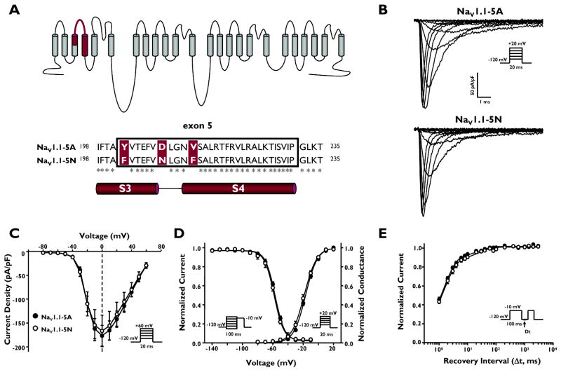
Figure 1.
Alternative splicing of SCN1A exon 5 results in two splice variants, NaV1.1-5A and NaV1.1-5N. (A) Predicted transmembrane topology of NaV.1.1 showing the location of the exon 5 coding region and amino acid alignment of NaV1.1-5A and NaV1.1-5N. Asterisks indicate identical amino acids. (B) Whole-cell sodium currents recorded from tsA201 cells co-expressing either NaV1.1-5A or NaV1.1-5N with both β1 and β2 accessory subunits. Channels were activated by voltage steps to between −80 and +60 mV from a holding potential of −120 mV. (C) Peak current density elicited by test pulses to various membrane potentials and normalized to cell capacitance. (D) Voltage dependence of channel activation measured between −80 to +20mV and voltage dependence of inactivation measured following a 100 ms inactivating prepulse to between −140 and −10 mV. (E) Time-dependent recovery from inactivation assessed with a 100 ms inactivating prepulse to −10 mV. Closed symbols represent NaV1.1-5A and open symbols represent NaV1.1-5N.

