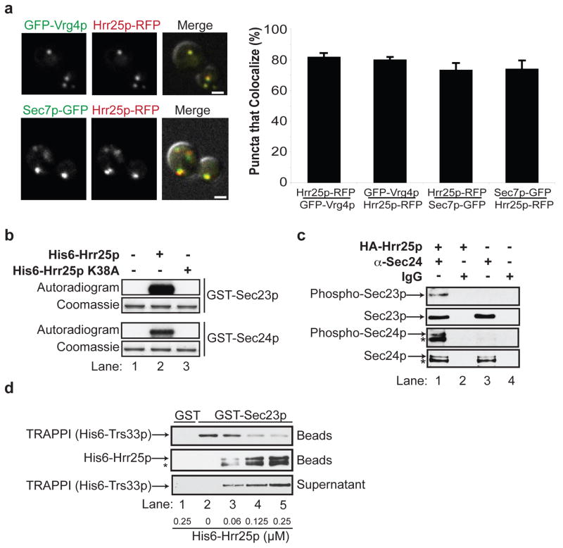
Figure 2. Hrr25p resides on the Golgi and phosphorylates Sec23p/Sec24p.
a, Hrr25p-RFP colocalizes with GFP-Vrg4p (top) and Sec7p-GFP (bottom). The green and red channels are merged with the DIC image (right panel). The puncta that colocalize (S.D., N=3) are shown on the right. The scale bar is 2 microns. b, GST-Sec23p and GST-Sec24p were incubated without (lane 1), or with His6-Hrr25p (lane 2) or His6-Hrr25p-K38A (lane 3) and γP32-ATP. The autoradiogram and coomassie stained gel are shown. c, Lysates expressing (lanes 1 and 2) or not expressing HA-Hrr25p (lanes 3 and 4) were immunoprecipitated with anti-Sec24 antibody (lanes 1 and 3) or IgG (lanes 2 and 4) and immunoblotted with anti-phosphoSer/Thr, anti-Sec23p and anti-Sec24p antibodies. d, TRAPPI, pre-bound to GST-Sec23p-containing beads (top panel), was incubated with increasing concentrations of His6-Hrr25p. The beads were pelleted and the amount of TRAPPI in the supernatant (bottom panel) and pellet (top panel) was assessed. The Hrr25p that bound to the beads (middle panel) was also measured. TRAPPI, or Hrr25p, did not bind to GST (lane 1). The starred bands in c and d are degradation products of Sec24p and His6-Hrr25p.

