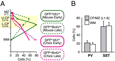
Fig. 3.
A majority of chicken MGE cells failed to enter the CP/MZ regardless of their birthdates and interneuron subtypes. (A) Quantification of the distribution of GFP-positive IdU-postive cells (green line), GFP-positive BrdU-postive cells (broken green line), GFP-negative IdU-postive cells (magenta line), and GFP-negative IdU-postive cells (broken magenta line) in the neocortical layers at P7. The percentages of the numbers of the four differentially labeled cells in each layer were analyzed (mean ± SEM; n = 2 brains, 59 slices, 226 GFP-positive IdU-positive cells, 81 GFP-positive BrdU-positive cells, 221 GFP-negative IdU-positive cells, and 349 GFP-negative BrdU-positive cells). Transplantation was performed as illustrated in Fig. S5A. (B) Quantification of the expression of MGE-derived interneuron subtype markers in mCherry-expressing chicken MGE cells in the CP/MZ (layers 1–6) or white matter (mean ± SEM; n = 7 brains for all analyses, 122 mCherry cells in the CP/MZ for PV analysis, 213 cells in the white matter for PV analysis, 85 cells in the CP/MZ for SST analysis, and 187 cells in the white matter for SST analysis). There were no statistically significant differences between the CP/MZ (layers 1–6) and the white matter in the percentages of mCherry cells that were PV-positive (P = 0.805, Mann–Whitney u test) or SST-positive (P = 0.906). Transplantation with chicken MGE cells was performed as illustrated in Fig. S2.

