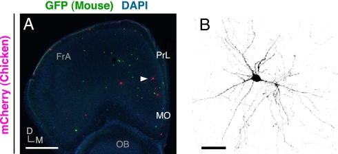
Fig. 4.
The neocortical CP is essentially permissive in regard to the postmigratory development of chicken MGE cells. (A) Distribution of GFP-expressing mouse MGE cells (green) and mCherry-expressing chicken MGE cells (magenta) in a coronal section of host forebrain at 4 wk after transplantation. Transplantation was performed directly into medial prefrontal cortex of P0 neonatal mice. (B) Enlarged view of the cell indicated by the arrowhead in A. FrA, frontal association cortex; PrL, prelimbic cortex; MO, medial orbital cortex; OB, olfactory bulb; D, dorsal, M, medial. (Scale bars: A, 600 μm; B, 25 μm.)

