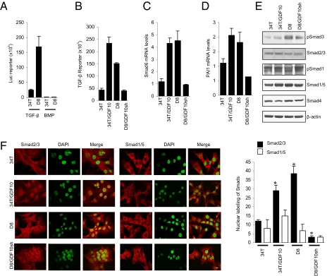
Fig. 2.
Silencing Sca-1 expression activates GDF10-dependent signaling through Smad3. (A) D8 cells exhibit increased Smad3 (TGF-β), but not Smad1/5 (BMP) reporter activity. D8 cells expressed a >20-fold increase in Smad3 activity compared with 34T cells. (B) GDF10 expression increases Smad3 activity. Reporter activity was increased in GDF10-overexpressing 34T cells (34T/GDF10), whereas silencing GDF10 expression in D8 cells (D8/GDF10sh) reduced the activity. (C and D) Expression of the TGF-β target genes Smad6 (C) and PAI1 (D) is regulated by GDF10 expression. Both Smad6 and PAI1 expression were increased by overexpression of GDF10 in 34T cells and decreased by down-regulation of GDF10 expression in D8 cells. (E) GDF10 expression regulates Smad3 activation, as assessed by immunoblotting for phosphoSmad3 (pSmad3). The levels of Smads and pSmads were determined by Western blot analysis. pSmad3 was increased in 34T/GDF10 cell, compared with 34T cells and was reduced in D8/GDF10sh cells compared with D8 cells. No difference in pSmad1/5 was observed. (F) GDF10 expression correlates with Smad2/3 nuclear localization. Fluorescence was measured by confocal microscopy using a Smad2/3 or Smad1/5 antibody and an Alexa Fluor 594-conjugated secondary antibody; nuclei were stained with DAPI and pseudo-colored in green. The merged image shows nuclear localization of Smad2/3 in 34T/GDF10 and D8 cells, but not in 34T and D8/GDF10sh cells. The bar graph quantifies Smad2/3 and Smad1/5 nuclear localization. *P < 0.01, Student two-tailed t test. (Scale bar: 10 μm.)

