Fig. 5.
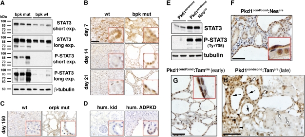
STAT3 is activated in PKD. Immunoblots of kidney tissue lysates from 21-d-old wild-type and bpk mutant mice (A) or 49-d-old Pkd1cond/cond and Pkd1cond/cond:Nescre mice (E). All other panels: Kidney sections derived from (B) bpk mutant mice, (C) orpk;Tg737Rsq mice, (D) human ADPKD kidney, (F) 49-d-old Pkd1cond/cond:Nescre mice, (G) tamoxifen-treated (P12) 21-d-old Pkd1cond/cond:Tamcre mice, and (H) tamoxifen-treated (P14) 6-mo-old Pkd1cond/cond:Tamcre mice were subjected to immunostaining for pY-STAT3. Representative fields and age-matched controls are shown. (H) Note that pY-STAT3 is highly expressed in cyst-lining cells (black arrows) and normal-appearing tubules (black arrowheads) and interstitial cells (white arrowheads) in close proximity to cysts, whereas normal tubules at a distance from cysts are negative (white arrows). (B–D) (Scale bars, 100 μm.) (F–H) (Scale bars, 50 μm.)

