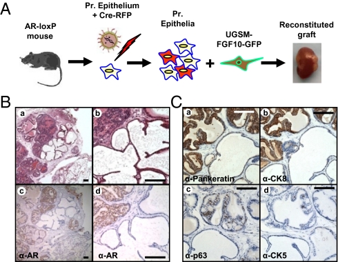
Fig. 4.
Loss of cell autonomous AR in adult prostate epithelium did not alter the tumorigenic capacity of FGF10-overexperssing stromal microenvironment. (A) The 105 dissociated prostate epithelia harvested from ARflox/Y mice were infected with a Cre lentiviral vector resulting in the loss of AR in a subset of epithelia. These epithelia were combined with 2 × 10⁵ FGF10-UGSM and were regenerated. (B) Areas of hyperplasia were seen adjacent to atrophic appearing glands within the same regenerated grafts (a and b). Immunohistochemistry revealed high levels of AR in the hyperplastic glands and absence of AR detection in the atrophic prostate glands (c and d). The normal prostatic secretions seen in the hyperplastic glands was absent in the atrophic tubules. (C) AR-null regenerated tubules were epithelial because they expressed pancytokeratin (a). These atrophic glands expressed low levels of prostate luminal marker CK8 (b) and had a diminution in number of basal cells compared with adjacent control/hyperplastic glands (c and d). (Scale bars: 100 μm.)

