
Figure 1.
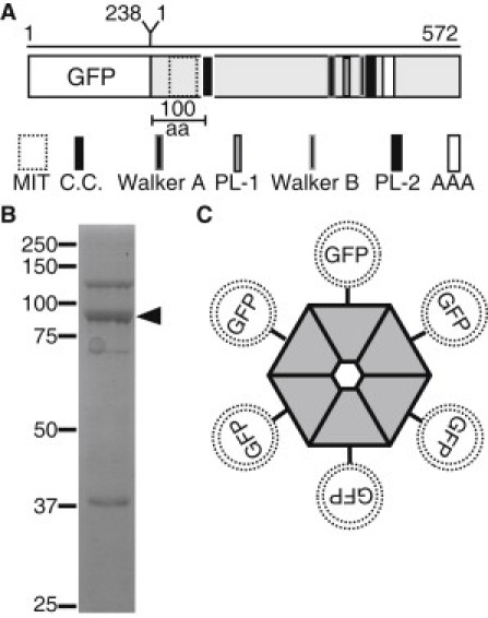
Description of domain architecture and purification of GFP-Katanin-60 from Sf9 insect cells. (A) Schematic depiction, approximately to scale, of domain architecture of GFP-Katanin-60. The amino acid length of GFP and Katanin-60 are indicated at the top of the figure. (aa, amino acids; MIT, microtubule interaction and trafficking domain; C.C., coiled-coil; PL-1, Pore Loop-1; PL-2, Pore Loop-2; AAA, AAA minimum consensus ATPase domain, Walker A, Walker B) (B) A coomassie stained SDS-PAGE gel of purified 6×His-tagged GFP-Katanin-60 after protein purification. Arrow marks the position of GFP-Katanin-60 (92 kD) that is the major band and molecular mass markers in kDa indicated at the left. (C) Cartoon of hexameric GFP-Katanin-60 ring showing the location of GFP at NH-terminal.