
Figure 2.
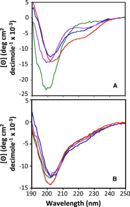
(A) CD spectra of the linker helix peptides of p27, p21, p57, and LH3G. Each sample contained 60 μM peptide in a buffer containing 20 mM sodium phosphate, pH 7.0 and 1 mM DTT. The spectra for p27, p21, p57, and LH3G linker helix domain peptides are red, green, blue, and purple, respectively. (B) CD spectra of the p27-KIDwt and the LH subdomain variants. Each sample contained 20 μM protein in the same buffer used for the linker helix peptides. Spectra were recorded at 25°C. The spectra for p27-KIDwt, p27-KIDp21LH, p27-KIDp57LH, and p27-KIDLH3G are red, green, blue, and purple, respectively.