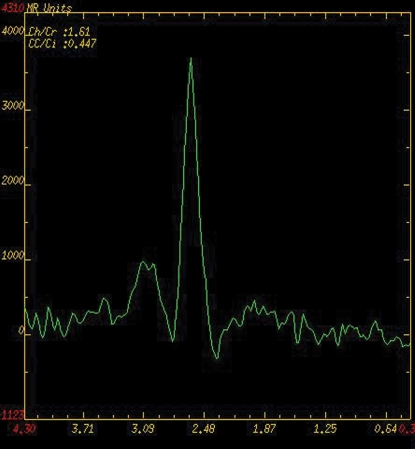
Figure 9a.
Comparison of MR spectroscopic spectra obtained in the peripheral zone of the normal prostate in different patients. (a) MR spectroscopic spectrum, obtained at 1.5 T with an endorectal coil, shows a high citrate (Ci) peak (resonance at 2.6 ppm) and a low choline (Ch) peak (resonance at 3.2 ppm), characteristics of benign tissue. The choline and creatine (Cr) peaks are overlapping. Ch + Cr/Ci = 0.447. (b) MR spectroscopic spectrum, obtained at 3 T with a body matrix coil, shows good separation of the choline (Cho) and creatine (Cr) peaks at higher magnetic field strength. The spectrum is normal, with a high concentration of citrate (Ci) and low concentration of choline. A diagnostic high-quality spectrum could be obtained with a noninvasive MR procedure without use of an endorectal coil. (c) MR spectroscopic spectrum, obtained at 3 T with an endorectal coil, shows a normal spectrum with a high citrate (Ci) peak and low choline (Cho) peak. Note the excellent spectral resolution of the citrate, choline, and creatine (Cr) metabolites, with only minimal baseline noise and detailed morphology of the citrate peak, which shows the effect of increased spectral resolution at 3 T, leading to a dominant central peak at 2.6 ppm with symmetric side peaks.

