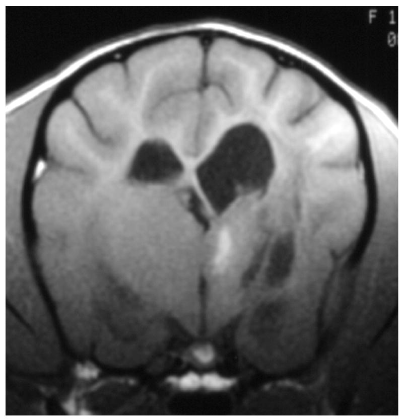
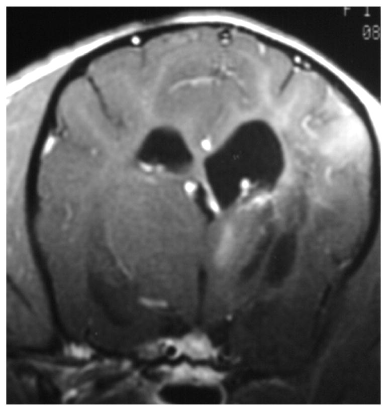
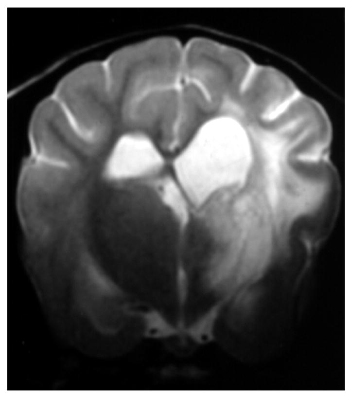
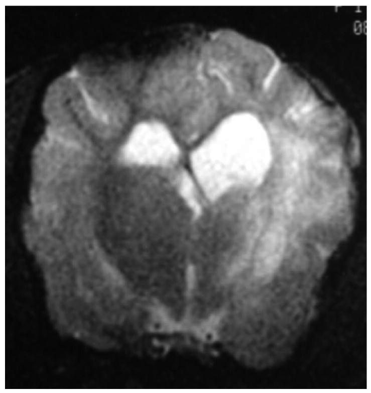
Figure 1.
Images of the brain of a 1 year old Maltese dog with necrotizing encephalitis. Extensive signal abnormalities are present throughout the gray and white matter of the left cerebrum and thalamus. Mild to moderate gadolinium enhancement is present in the cerebrum and overlying meninges. Focal parenchymal loss and replacement with fluid are visible in the left thalamus and temporal lobe. (A - T1-weighted image; B – T1-weighted image + gadolinium; C – T2-weighted image, D – gradient echo image)


