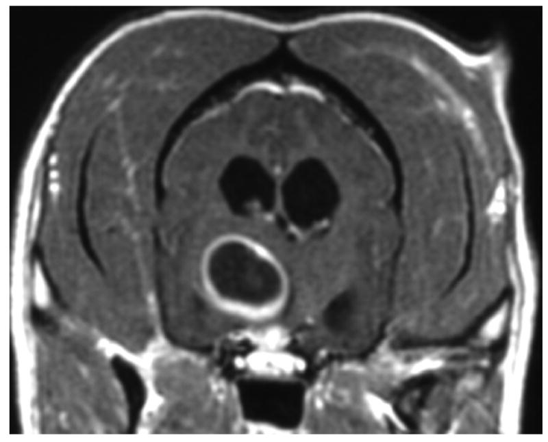
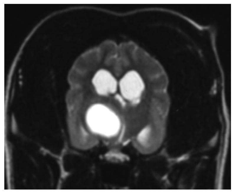
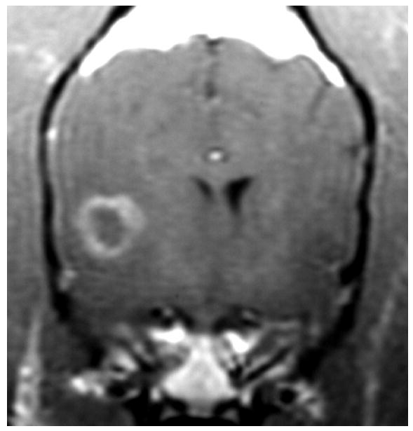
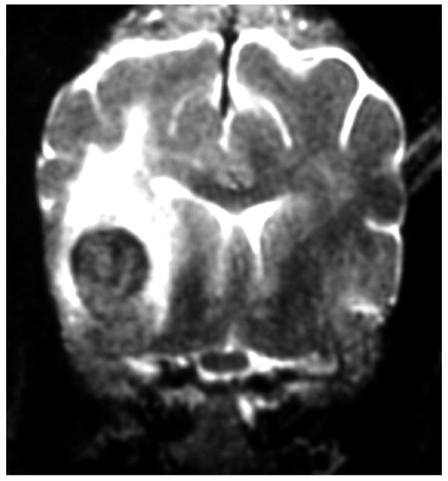
Figure 4.
Images A and B are of the brain of an 8 year old mixed-breed dog with an oligodendroglioma within the right side of the thalamus. The lesion shows strong ring-enhancement and the presence of a central cavity with signal characteristics distinct from that of CSF. Images C and D are of the brain of a 14 year old mixed breed dog with an infarct within the right temporal lobe. The lesion shows moderate to strong ring-enhancement, regionally extensive edema, and evidence of hemorrhage. (A & C – T1-weighted image + gadolinium; B &D – T2-weighted image)


