Figure 5.
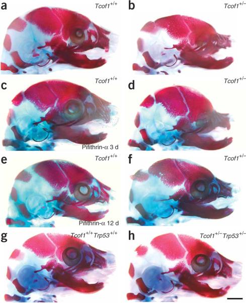
Embryonic skeletal analyses and prevention of TCS. Craniofacial skeletons of E17.5 embryos were stained for bone (alizarin red) and cartilage (alcian blue). (a) Wild-type Tcof1+/+ embryo, showing a normal craniofacial skeleton. (b) Mutant Tcof1+/− embryo, showing severe frontonasal hypoplasia including the nasal, frontal, premaxilla, maxilla and mandibular bones, and doming of the skull and severe microphthalmia. (c) Tcof1+/+ embryo treated with pifithrin-α for 3 d in utero, showing a normal craniofacial skeleton. (d) Tcof1+/− embryo treated with pifithrin-α for 3 d in utero, showing slightly reduced frontonasal hypoplasia and less severe microphthalmia. (e) Tcof1+/+ embryo treated with pifithrin-α for 12 d in utero, showing a normal craniofacial skeleton. (f) Tcof1+/− embryo treated with pifithrin-α for 12 d in utero, showing substantially reduced frontonasal hypoplasia with considerable restoration of normal patterning of the frontal, nasal, premaxilla, maxilla and mandibular bones, and little evidence of any persistent calvarial doming or microphthalmia. (g) Wild-type Tcof1+/+Trp53+/+ embryo, showing a normal craniofacial skeleton. (h) Tcof1+/−Trp53+/− littermate embryo, showing a completely normal craniofacial skeleton that is indistinguishable from wild-type. Scale bar, 2 mm.

