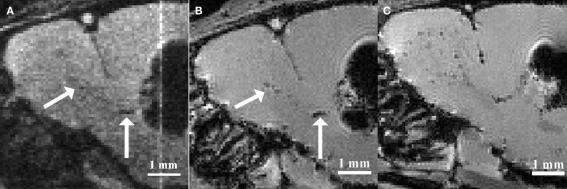
Figure 1.
Magnetic resonance imaging of in situ labeled neural precursor cell migration. (A) In vivo MRI of a mouse injected with micron-sized iron oxide particles in the lateral ventricle at 3 weeks post-injection. (B) Ex vivo MRI of the same mouse at 3 weeks post-injection. (C) Ex vivo MRI of a mouse injected with micron-sized iron oxide particles in the lateral ventricle at 8 weeks post-injection. Arrows indicate corresponding hypointense contrast on the in vivo and ex vivo MRI of the same mouse (Adapted and reproduced from Vreys et al., 2010).

