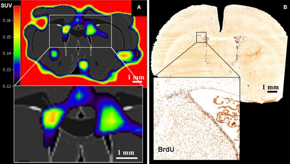
Figure 3.
Positron emission tomography imaging of neural stem cell proliferation ([18F]-FLT–PET matched on MRI-atlas of rat brain). (A) Increased [18F]-FLT signal was detected in the SVZ. (B) Location of BrdU-labeled proliferating cells in the SVZ corresponds well with the elevated [18F]-FLT signal in the SVZ (A) (Adapted from Rueger et al., 2010).

