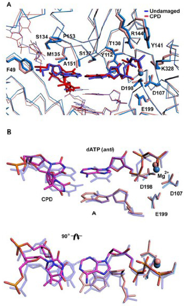
Figure 3.
Comparison between T-T dimer and undamaged DNA ternary Polκ complexes. (A) Superimposition of the T-T dimer complex (red) and the complex with undamaged DNA (blue; PDB code 2OH2) (blue). Highlighted are the template bases (T-T dimer and A), incoming dNTPs (dATP and dTTP) and residues within the active site cleft. The putative Mg2+ ion is shown as a sphere in each structure. (B) Close-up views of the superposition of the T-T dimer (magenta) and undamaged DNA (blue), and the incoming nucleotides dATP (magenta) and dTTP (blue), respectively. The figure also shows the catalytic triad and the putative Mg2+ ion in each structure. The figure shows that the active site geometry is conserved in the damaged and undamaged structures.

