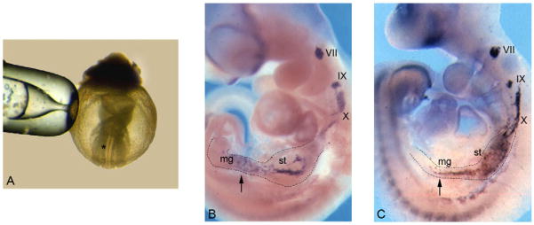
Fig. 1. Normal development of the peripheral nervous system in cultured embryos.
E8.5 mouse embryos (A) were cultured for 3 days and processed for whole-mount in situ hybridisation with a Ret-specific riboprobe (B). In situ hybridisation of a freshly dissected E10.5 embryo (C). In both cases, Ret expression is observed in the VII, IX and X cranial sensory ganglia and in the stomach (st) and midgut (mg). The asterisk in A marks the approximate site of cell grafting (see text, and legend to Fig. 2). Arrows indicate enteric neural crest cells (NCCs) within the gut of cultured (B) and freshly dissected (C) embryos.

