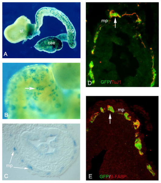
Fig. 3. Ret+ cells grafted to the vagal NCC pathway of E8.5 embryos are capable of colonising the entire length of the gastrointestinal tract.
(A) Whole-mount X-Gal staining of gut from E8.5+3 mouse embryos transplanted with Ret+ cells from the intestine of E11.5 Rosa26bgeo embryos. Note the efficient colonisation of the entire gut by β-geo+ cells. (B) Higher magnification of the gut shown in A. Arrow points to a cluster of β-geo+ cells. (C) Cryosections of E8.5+3+7 gut show that the progeny of grafted cells are localised within the myenteric plexus (mp, arrows). (D,E) Similar cryosections from embryos transplanted with YFP-expressing Ret+ cells were double immunostained for GFP and TuJ1 (D) or for GFP and B-FABP (E). st, stomach; mg, midgut; cae, caecum.

