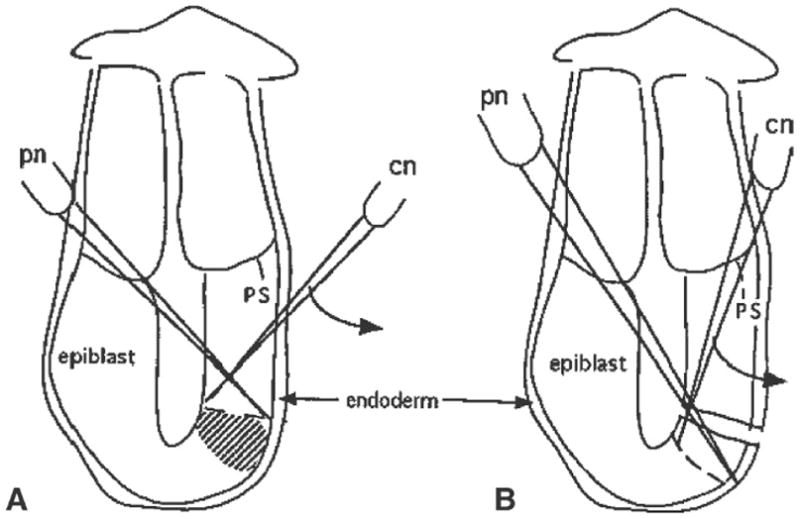
Fig. 2.

The steps taken to isolate epiblast fragments from sites adjacent to the distal cap region on the posterior side of a 6.5-d embryo. A Position of the pinning needle (pn) and the line of cut that will be made by the cutting needle (cn) just proximal to the required tissues (shaded area). B Second cut made to isolate the tissue fragment. Similar cutting actions are employed to isolate tissue fragments from other regions of the embryo. Curved arrows indicate the direction of slicing made by the cutting needle. Abbreviation: ps, primitive streak.
