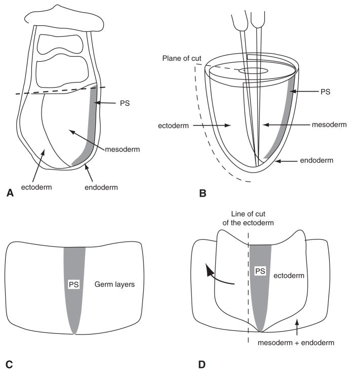
Fig. 3.
The steps of germ layer separation of 7.0–7.5-d embryos. A Tissue organization of the 7.5-d embryo. The dashed line marks the position of the first cut at the junction between the embryonic and extraembryonic parts of the egg cylinder. B The embryonic portion of the embryo that reveals the anatomical relationship of the three germ layers. A metal needle is pushed into the amniotic cavity and pins the egg cylinder in the upright position. The second metal needle is then brought inside the amniotic cavity, and the embryo is cut along the anterior side (indicated by the dashed line) by slicing the second needle through all germ layers. C The embryo opened out flat and ready for enzyme digestion. D The ectoderm layer that has been loosened by enzymatic digestion. The ectoderm can be lifted up and torn away from the mesoderm using needles. It is not possible to separate the germ layers at the site of the primitive streak. The ectoderm is therefore cut along the dashed line, where no further separation from the underlying primitive streak could be made. The mesoderm layer can be separated from the endoderm in the same manner, followed again by cutting them close to the primitive streak. The remaining tissue is the primitive streak (PS).

