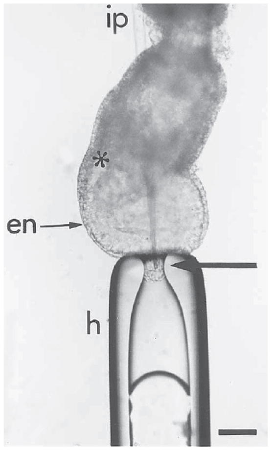
Fig. 7.

Labeling of the epiblast by microinjecting DiI into the distal cap of the embryo. To inject in the midline, the embryo is held at the distal tip of the egg cylinder by gentle suction with the holding pipet (h). The injection pipet (ip) is passed through the extraembryonic tissues of the egg cylinder. The injection pipet is brought to the site of labeling from within the pro-amniotic cavity to avoid inadvertent labeling of other embryonic germ layers. The arrow points to the tip of the labeling pipet in the epiblast layer: en, primitive endoderm; *primitive streak. Bar = 20 μm.
