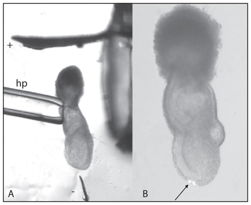
Fig. 8.

Labeling the endoderm of 7.0-d embryo by electroporation. A The embryo is first bathed in plasmid DNA solution before transfer to the electroporation drop on the culture dish. To electroporate the distal tip, the embryo is held at the extraembryonic ectoderm by suction using the holding pipet (hp) and positioned between the platinum plate (cathode) and tungsten needle (anode). The electrodes are connected to an Electro Square Porator and the electroporation is focused at the tissue closest to the needle tip. B Fluorescent cells in the distal region of the embryo following electroporation with a β-actin-eGFP construct and cultured for 3 h.
