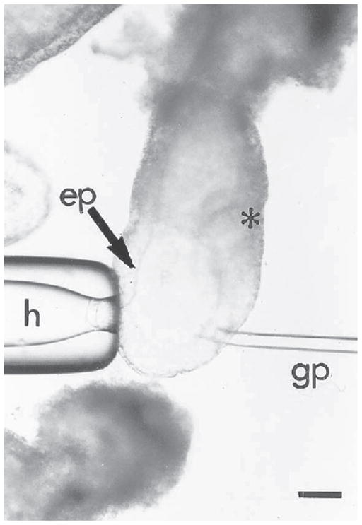
Fig. 9.

Grafting of cells to a 6.5-d embryo. The embryo is held on the anterior side opposite the intended site of grafting by the holding pipet (h). The cells are grafted into the epiblast (ep) on the posterior side of the embryo (*marks the position of the primitive streak) immediately proximal to the margin of the distal cap. Bar = 20 μm.
