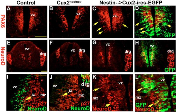
Fig. 3. Cux2 regulates neural progenitors and neuroblasts.
(A–D) Pax6 expression in spinal cords of E11.5 wild-type (A), Cux2neo/neo (B) and Nestin-Cux2-ires-EGFP (C,D) embryos. (D) GFP (green) overlay on anti-Pax6 immmunohistochemistry (red) revealed a mediolateral expansion of Pax6-labeled vz cells following Cux2 overexpression (arrows). (E–H) Neurod immunostaining of neuroblasts exiting the vz and also cells in the drg in E11.5 control (E), Cux2neo/neo (F) and Nestin-Cux2-ires-EGFP (G,H) spinal cords. (I,J) Anti-Neurod1 (green) labeling of neuroblasts in the iz (arrow) adjacent to anti-p27Kip1 (red) immunohistochemistry in E10.5 ventral spinal cords of control and Cux2neo/neoembryos. (K,L) Enhanced Neurod1 (red) activity (arrows) in an E10.5 Cux2 transgenic embryo overexpressing Cux2-ires-EGFP (green). Scale bars: 250 μm in A,E; 500 μm in I.

