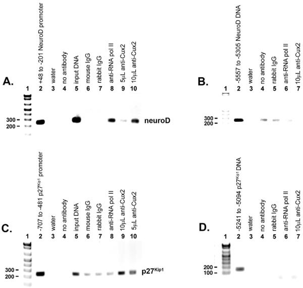
Fig. 5. Neurod1 and p27Kip1 are direct Cux2 targets in the embryonic nervous system.
Complexes isolated from E12.5 mouse brains were immunoprecipitated with either anti-Cux2 or anti-RNA polymerase II antibodies followed by reverse cross-linking of protein and DNA, and PCR amplification of a 248 bp fragment of the Neurod1 promoter spanning +48 to −201 (A), or a 227 bp fragment of the p27Kip1 promoter spanning −707 to −481 (C), relative to the transcription start sites. (A) Inverse image of ethidium-stained agarose gel showing PCR amplification of a 248 bp Neurod1 product from genomic DNA (lane 2), input DNA (lane 5), following ChIP with antibody directed against RNA polymerase II (lane 8), indicating that the Neurod1 gene is transcriptionally active, and following ChIP with 5 μl (lane 9) or 10 μl (lane 10) of anti-Cux2 antibodies, indicating that Cux2 interacts with the Neurod1 promoter in vivo. (B) To control for non-specific chromatin immunoprecipitation, a 250 bp product from −5557 to −5308 relative to the Neurod1 transcription start site was amplified from input DNA (lane 2), following ChIP with an anti-RNA polymerase II antibody (lane 6), and following ChIP with 10 μl (lane 7) of anti-Cux2 antibodies. (C) PCR amplification of a 227 bp p27Kip1 product from genomic DNA (lane 2), input DNA (lane 5), following ChIP with anti-RNA polymerase II antibodies (lane 8), indicating that p27Kip1 is transcriptionally active, and following ChIP with 10 ml (lane 9) or 5 ml (lane 10) of anti-Cux2 antibodies, indicating that Cux-2 interacts with the p27Kip1 promoter in vivo. (D) To control for non-specific chromatin immunoprecipitation, a 148 bp product from −5241 to −5094 relative to the p27Kip1 transcription start site was amplified from input DNA (lane 2), following ChIP with anti-RNA polymerase II antibodies (lane 6), and following ChIP with 10 μl (lane 7) of anti-Cux2 antibodies. In both A and C, controls were water (lane 3), no antibody (lane 4), normal mouse IgG (lane 6) and normal rabbit IgG (lane 7). In both B and D, controls were water (lane 3), no antibody (lane 4) and normal rabbit IgG (lane 5).

