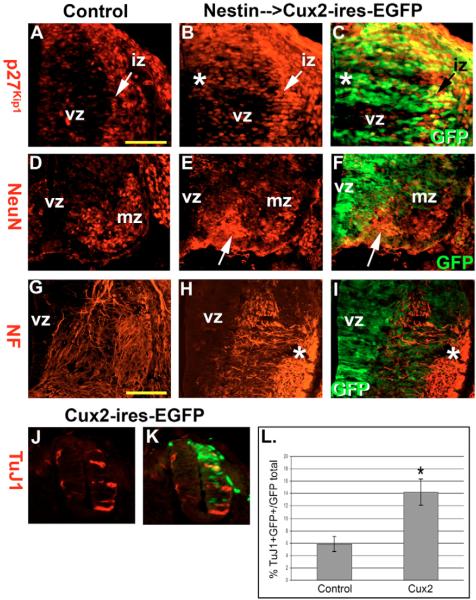
Fig. 6. Cux2 overexpression.
(A–C) P27Kip1 levels (red) in E11.5 wild-type (A) and Nestin-enhancer-driven Cux2-ires-EGFP (green) transgenic (B–C) spinal cords in which Cux2 enhances p27Kip1 in the vz and iz in a cell-autonomous manner (*). (D–F) NeuN immunohistochemistry on E11.5 control (D) and Cux2 transgenic (E,F) ventral spinal cords revealed that Cux2 increases NeuN-labeled post-mitotic neurons emerging from the vz (arrow). (G–I) Neurofilament immunohistochemistry labels axons growing towards the ventral midline in control embryos; however, in Cux2-ires-EGFP transgenic embryos, Cux2 overexpression induced ectopic disorganized and misdirected axons throughout the mz (*). (J,K) TuJ1 staining of HH stage 14–15 chick embryos electroporated with a murine Cux2-ires-EGFP vector. (L) Quantification of TuJ1+ and GFP+ double-positive cells following control vector or Cux2 overexpression. *P=0.05 by Student's t-test. Scale bars: 250 μm in A; 300 μm in G.

