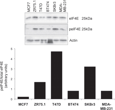
Figure 2.
eIF4E expression and phosphorylation in breast cancer tissue. Cells in exponential growth phase were lysed 24 h after the application of fresh medium. Proteins were separated by SDS-PAGE and western blots probed with antibodies to MNK1, eIF4E and serine 209 phosphorylated eIF4E. Figure representative of two independent blots. Quantitation is as per Figure 1.

