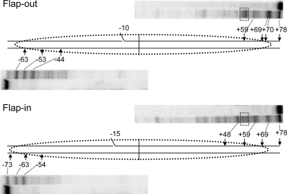
FIGURE 3.
ExoIII analysis of flap-out and flap-in 154-bp 5 S nucleosome substrates. Nucleosomes were purified and digested with ExoIII as described in the text, and cleavage products were analyzed by sequencing gels and phosphorimagery. Shown are lanes corresponding to undigested and digested samples for each 3′ end of the flap-out (FO, top) and flap-in (FI, bottom) substrates. Numbers indicate the distance from the expected nucleosome dyad fixed at position 0.

