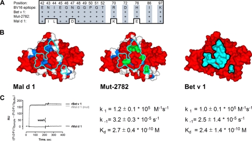
FIGURE 2.
A, amino acid sequence alignment of residues present within the epitope defined by the binding of the mAb BV16 to Bet v 1 in Bet v 1, Mal d 1, and the mutated Mal d 1 variant Mut-2782. Five residues within the antibody footprint are not shared between Mal d 1 and Bet v 1. All 16 residues within the antibody footprint are shared between Bet v 1 and Mut-2782. B, modeled molecular surfaces of Mal d 1, Mut-2782 with the BV16 epitope grafted onto its surface, and Bet v 1. Amino acid residues are color-coded according to amino acid identity (red) to Bet v 1 and (blue) indicates naturally occurring conservative substitutions. Right model shows Bet v 1 (Z80104) with 16 amino acid residues colored cyan that make up the BV16 epitope. Left model shows Mal d 1 (Q8L6K9) with the antibody footprint visible in cyan. Center model shows Mut-2782 with the antibody footprint visible in cyan and mutated amino acid residues colored in green. C, kinetic parameters for the binding of mAb BV16 are shown below each model. No binding to Mal d 1 could be detected, whereas very similar kinetic parameters characterize BV16 antibody binding to Bet v 1 and Mut-2782 were found.

