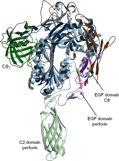
FIGURE 6.
Comparison of C8 and perforin structures. C8 is in dark colors and perforin in light colors. C8γ is in green, and C8β is omitted. The MACPF/CDC domains are in blue, EGF domains in purple, and remaining domains in brown. The putative membrane location would be horizontal and below the models.

