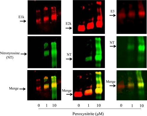
FIGURE 3.
Western blots of KGDHC separated by Blue-native gel. Purified KGDHC was treated with different concentrations of peroxynitrite, and subjected to a Blue-native gel followed by Western blotting. The blots were probed with antibodies to E1k, E2k or E3 (red) and to nitrotyrosine (green), respectively. The overlap is represented by yellow.

