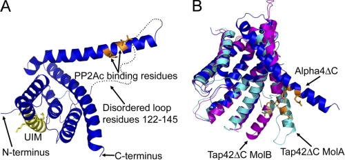
FIGURE 1.
Structure of Alpha4ΔC. A, ribbon diagram of Alpha4ΔC, with residues important for PP2Ac binding shown in orange and the consensus UIM shown in yellow. B, comparison of the Alpha4ΔC structure (blue) with the Tap42ΔC structures (cyan and magenta) showing the variable positions of the extended helix (residues 147–182). PyMOL was used to depict all molecular structures (48). MolA and MolB, molecules A and B, respectively.

