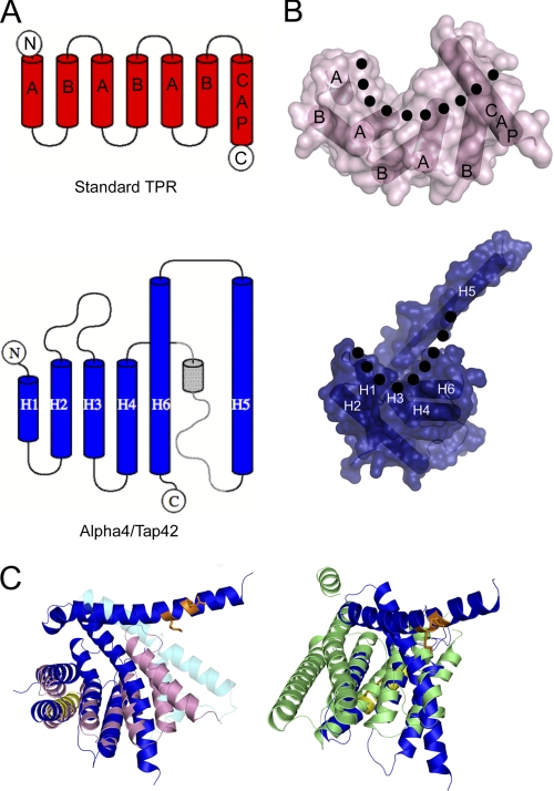
FIGURE 3.
Comparison of Alpha4ΔC and TPR proteins. A, topology diagrams of TPR (upper) and Alpha4ΔC (lower) showing the altered topology of the final helices. The part represented in gray is based on the crystal structure of Tap42ΔC, as these residues are not observed in the crystal structure of Alpha4ΔC. The diagrams were created in TOPDRAW (49). B, structures and surface representations of TPR (upper) and Alpha4ΔC (lower) showing the configuration of helices and the formation of the concave and convex surfaces (with the outline of concavity denoted by the dotted line). C, superposition of Alpha4ΔC (colored blue, yellow, and orange as in Fig. 1) with the SycD TPR domain (pink; Protein Data Bank code 2VGY) and 14-3-3 (green; code 3EFZ) reveals similar tertiary structures but indicates that the concave face of Alpha4 is more closed than the canonical TPR and 14-3-3 proteins. The helices shown in cyan represent helices from Tap42ΔC that differ significantly in position from those in Alpha4ΔC.

