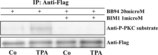
FIGURE 3.
TPA induces serine phosphorylation on NRG that can be blocked by PKC inhibition. MEF cells were preincubated with the metalloproteinase inhibitor batimastat (BB94, 20 μm) and with either control (DMSO) or with an inhibitor of classical PKC isoforms bisindolylmaleimide I (BIM1, 1 μm). Subsequently, cells were stimulated for 30 min with either control medium (DMSO) or TPA (1 μm). After stimulation, cells were placed on ice and incubated with anti-FLAG antibody (on plate), prior to cell lysis, to capture only the cell surface fraction of the overexpressed reporter FLAG-NRG-GFP. Anti-FLAG-NRG complexes were immunoprecipitated (IP) with protein G-agarose and resolved by SDS-PAGE with the antibodies indicated. Anti-P-PKC-substrate antibody recognizes phosphorylated serine within a consensus PKC phosphorylation site (arginine or lysine in position −2 and +2 and a hydrophobic amino acid at position +1 relative to the serine). Shown is one of four identical experiments with the same outcome.

