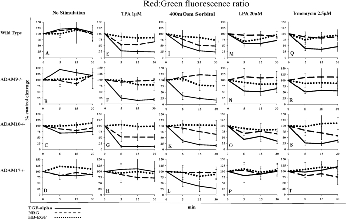
FIGURE 4.
Comparison of pro-EGF ligand cleavage in wild type MEFs and MEFs knock-out for either ADAM9, -10, or -17. Cleavage of pro-EGF reporter ligands was detected by changes in the cellular red:green fluorescence ratio as measured by FACS (details see text). Red:green fluorescence was plotted over time and compared with % control in wild type or ADAM knock-out mouse embryonic fibroblasts (lacking either ADAM9, -10, and -17) stably overexpressing precursors of either TGFα (solid lines), NRG (dashed lines), or HB-EGF (dotted lines). Plots for each different cell line are shown in horizontal rows, whereas plots for different cell lines subjected to the same cleavage stimulus are arranged in vertical columns. Cells were either control-treated (A–D) or incubated with TPA 1 μm (E–H), sorbitol 400 mosm (I–L), LPA 20 μm (M–P), or ionomycin 2.5 μm (Q–T) and monitored by FACS at 5, 15, and 30 min.

