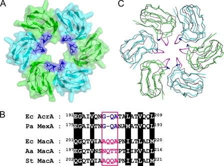
FIGURE 2.
Putative pore region of the AcrA hexameric model. A, central pore in the lipoyl domains from the AcrA hexamer. Only the lipoyl domains are shown for clarity. Each protomer is colored green or cyan with transparent surface representations. The Gly-Gln-Ala sequence conserved between E. coli AcrA and P. aeruginosa MexA is shown in blue. B, alignment of the sequences around the pore-lining loops from E. coli AcrA (Ec AcrA), P. aeruginosa MexA (Pa MexA), E. coli MacA (Ec MacA), A. actinomycetemcomitans MacA (Aa MacA), and Salmonella typhimurium MacA (St MacA). The pore-lining sequences are colored in blue or magenta in a box. The conserved residues are highlighted. C, superposition of the lipoyl domains from E. coli MacA and E. coli AcrA hexamers, displayed in the Cα tracing representations. The E. coli MacA protomers are colored in gray, although E. coli AcrA protomers are in green or cyan. The pore-lining residues of MacA are in magenta, and those of AcrA are in blue.

