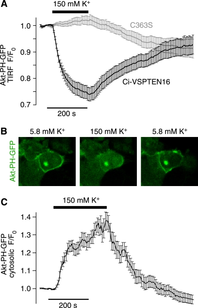
FIGURE 6.
Experimental control of Ci-VSPTEN activity in intact cells without use of electrophysiological instrumentation. A, reversible dissociation of Akt-PH from the plasma membrane upon K+-induced depolarization observed with Ci-VSPTEN16 (n = 30 cells from five independent experiments) but not with the catalytically inactive Ci-VSPTEN16-C363S (n = 29 cells, from five independent experiments), measured by TIRF microscopy. CHO cells were cotransfected with Ci-VSPTEN16, Akt-PH-GFP, PI3K, and the potassium channel TASK3. B, confocal images of OK cells show reversible translocation of Akt-PH from the plasma membrane to the cytosol upon K+-induced depolarization. OK cells were cotransfected as described in A. C, averaged time course of K+-induced translocation of Akt-PH-GFP obtained from experiments as described in B (n = 19 cells from two independent experiments).

