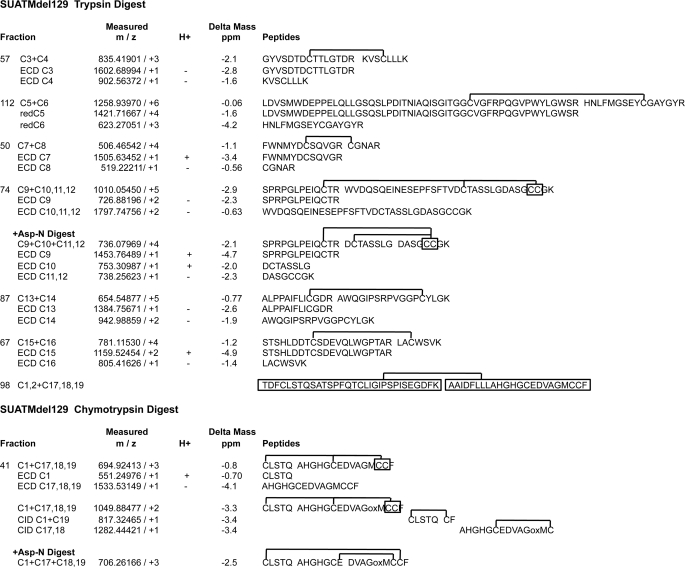
FIGURE 4.
Summary of the MS analysis of disulfide-bonded peptide pairs of SUATM129. The accurate mass measurements of the disulfide-bonded peptide pairs were made using FTICR mass spectrometry with an LTQ-FT. The measured masses are shown as mass/ion charge (m/z). ECD was used to preferentially break the disulfide bond of the peptide pair to yield the original peptide pair, and each peptide mass was measured (e.g. ECD C3; ECD C4). One difference to note between using ECD compared with chemical reduction of a disulfide bond: ECD does not necessarily result in peptides with replaced hydrogens on all of the now free sulfur atoms. Peptides generated by ECD that have (+) or have not (−) replaced the hydrogen ions are indicated in the H+ column. For peptide pair C5+C6, the fraction was reduced and analyzed by LTQ-FTICR MS to measure the masses of the reduced peptides (redC5; redC6). Peptide pairs from CID that define the disulfide bond pattern between cysteines C1 and C17,18,19 are shown with the complete data presented in Fig. 7. The C1, C2+C17, C18, C19 peptide pair was identified only by Edman chemical sequencing. The analytical difference between the observed mass compared with the theoretical mass is given as Delta Mass in parts per million. The peptide sequences corresponding to the measured mass are shown with the putative disulfide bonds. The SUATM129 glycoprotein contains two adjacent cysteine pairs: C11,C12 and C18,C19 (marked in boxes). The methionine in the C17, C18, C19 peptide in Fraction 41 was found to be oxidized (oxM) after freeze/thaw from storage: this was the predominant form in the Asp-N and CID analysis. We consistently observed an unusual chymotrypsin cleavage between Gln29 and Ser30 generating the C1 peptide CLSTQ in multiple digests. We have no definitive explanation, but it may reflect the extended overnight digestion at room temperature.

