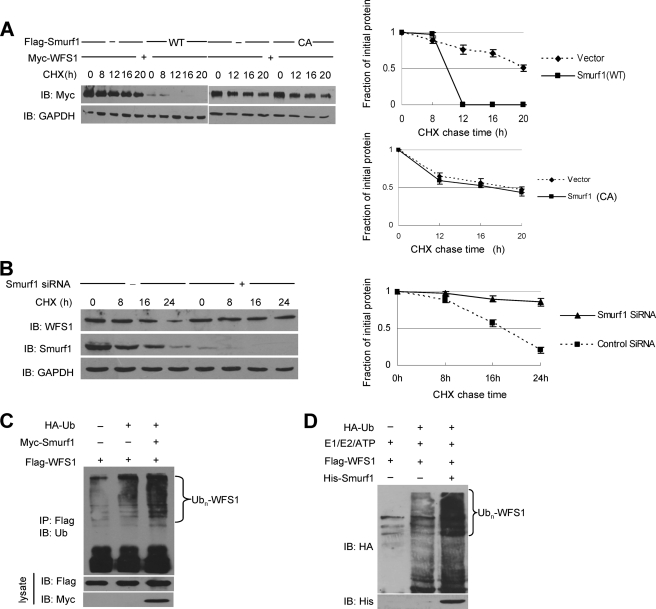
FIGURE 4.
Smurf1 promotes the ubiquitination and proteasomal degradation of WFS1. A, the effect of Smurf1 on the half-life of WFS1 is shown. Smurf1 WT or C699A (CA) was transfected together with a WFS1 expression vector, and cells were treated with CHX at 10 μg/ml for the indicated times. The half-life of WFS1 was measured by Western blot. B, CHX-chase experiments of WFS1 in MCF7 cells transfected with scrambled siRNA or Smurf1 siRNA are shown. Cells were treated with CHX at 10 μg/ml for the indicated times. The half-life of WFS1 was measured by a Western blot. C, Smurf1 promotes WFS1 ubiquitination in mammalian cells. HEK293T cells were transfected with HA-tagged ubiquitin (HA-Ub), FLAG-tagged WFS1, control vector, or Myc-tagged Smurf1. Sixteen hours before cell harvest, the cells were treated with the potent proteasome inhibitor lactacystin (30 μm) to avoid the proteasome-mediated degradation. Cell lysates were then prepared and immunoprecipitated with an anti-FLAG antibody. The immunoprecipitates (IP) were analyzed by immunoblotting with the anti-ubiquitin antibody to indicate the polyubiquitinated WFS1. The lysates were also analyzed by immunoblotting with anti-FLAG and anti-Myc antibodies. D, Smurf1 enhances WFS1 ubiquitination in vitro. E1, UbcH5c (E2), HA-Ub (all from Boston Biochem), His-Smurf1 (expressed in bacteria and purified), and FLAG-WFS1 (expressed in HEK293T cells and purified by immunoprecipitation with an anti-FLAG antibody) were incubated at 30 °C for 2 h in ubiquitination buffer. Ubiquitinated WFS1 was visualized by immunoblotting with an anti-HA antibody.

