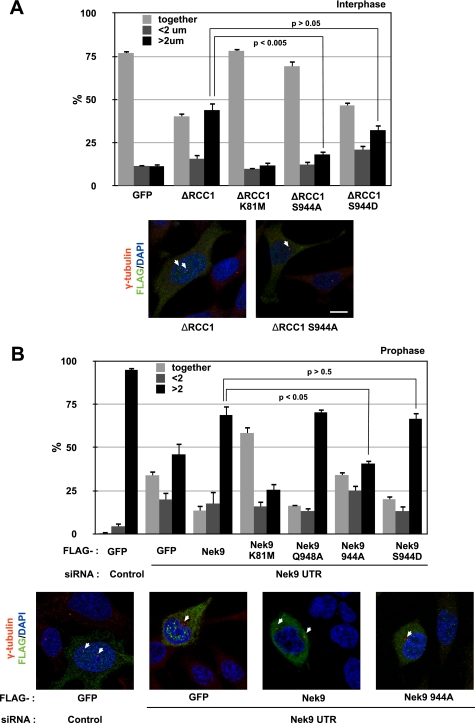
FIGURE 6.
LC8 binding to Nek9 regulates its physiological activity. A, exponentially growing HeLa cells transfected either with FLAG-GFP or with different FLAG-Nek9 [Δ346–732] (ΔRCC1) forms were fixed and stained with antibodies against FLAG and γ-tubulin to detect centrosomes and DAPI to visualize DNA. The percentages of FLAG-positive cells showing two unseparated centrosomes (together), two centrosomes separated less than 2 μm (<2 μm), and fully separated centrosomes (>2 μm) are shown in the upper panel (mean ± S.E. of three independent experiments; ∼50 cells were counted in each experiment, and statistical significance was determined using the standard Student's t test). Representative examples of the observed phenotypes are shown below (bar, 10 μm). B, exponentially growing HeLa cells were transfected with either control or Nek9 3′-UTR siRNAs, and 24 h later, they were transfected with expression plasmids for the indicated FLAG-tagged proteins. After an additional 24 h, cells were fixed and processed as in A. FLAG-GFP was used as a control protein. Prophase cells (showing condensed chromosomes and intact nuclei as assessed by the shape of the DNA and a γ-tubulin exclusion) were categorized according to centrosome separation (mean ± S.E. of three independent experiments; ∼50 cells were counted in each experiment, and statistical significance was determined using the standard Student's t test). Representative examples of the observed phenotypes are shown below (bar, 10 μm).

