Figure 4.

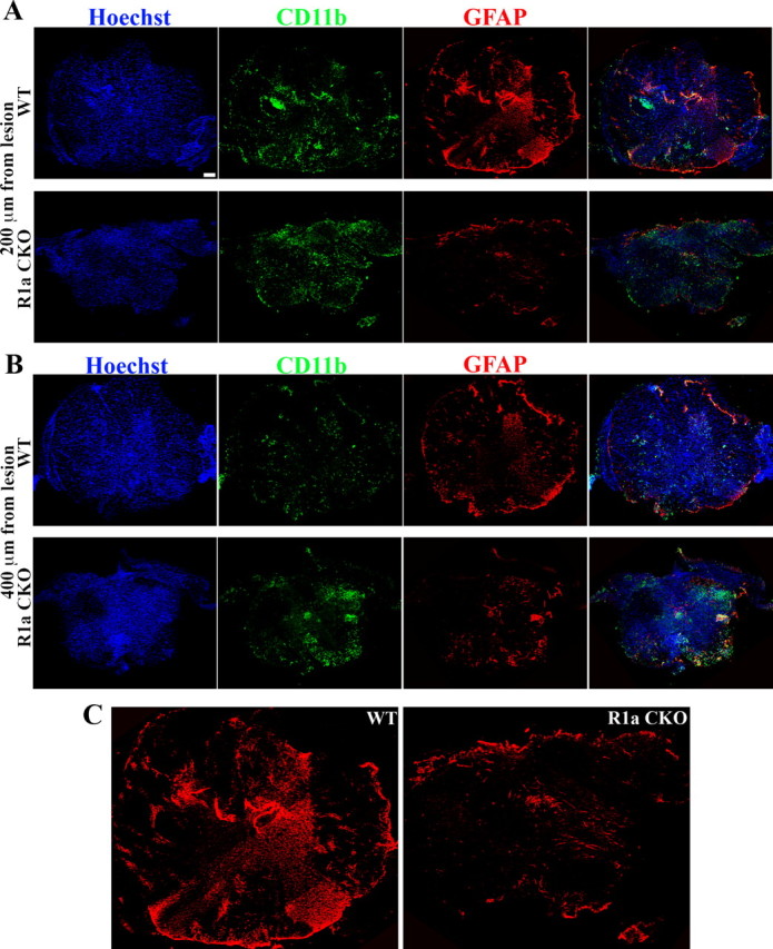
Reduction in GFAP area in BMPR1a CKO mice indicating defective gliosis is independent of the Cre line. A, Cross-sections taken at 200 μm distance from the lesion rostral to it from either GFAP Cre; BMPR1a +/+ (WT) animals or GFAP Cre; BMPR1a flox/− animals (BMPR1a CKO) at 1 week postinjury stained with Hoechst nuclear stain (blue), CD11b (green) and GFAP (red). B, Cross-sections similar to those from the genotypes in A at 400 μm distance from the lesion rostral to it. Scale bars (in A, B), 100 μm. C, Larger images of GFAP-stained sections shown in A showing reduced GFAP staining in the BMPR1a CKO compared with WT animals.
