Figure 5.

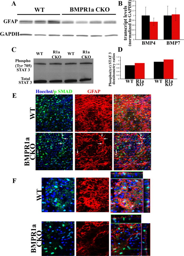
BMPR1a signaling affects GFAP levels independent of the canonical SMAD signaling pathway. A, Western blots for GFAP protein in injured cords from WT and BMPR1a CKO mice at 6 d post-SCI. The blot was stripped and reprobed for GAPDH (loading control). B, Normalized transcript levels of BMP4 and BMP7 in cords from WT and BMPR1a CKO mice at 6 d post-SCI showing no significant difference between the groups. C, Western blot analysis for Phospho (Tyr) STAT3 and total STAT3 in injured cords from 2 sets of littermates of WT and BMPR1a CKO animals at 4 d post-SCI. Activated STAT3 is present in the BMPR1a CKO mice. D, Densitometric analysis of the individual littermate pairs showing a slight increase in the levels of phospho-STAT3 as normalized to total STAT3 in the cords from BMPR1a CKO animals. E, Confocal Z-stacks taken from WT and BMPR1a CKO injured spinal cords (6 d post-SCI) stained for Hoechst nuclear stain (blue), npSMAD (green) and GFAP(red). Defective reactive astrocytes in BMPR1a CKO mice still have robust npSMAD (arrows). Scale bar, 20 μm. F, Single confocal Z-sections (from the stacks shown in E) taken at higher magnification with orthogonal projections showing intact npSMAD staining in the BMPR1a CKO astrocytes.
