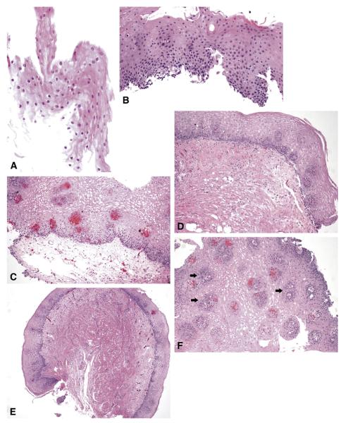
Figure 1.
A, Partial-thickness squamous epithelium. B, Full-thickness squamous epithelium. C, Squamous mucosa with abundant underlying LP. D, Squamous mucosa with underlying muscularis mucosae. E, Squamous mucosa with underlying muscle and small amount of submucosa. F, Squamous mucosa with numerous LP papillae (arrows).

